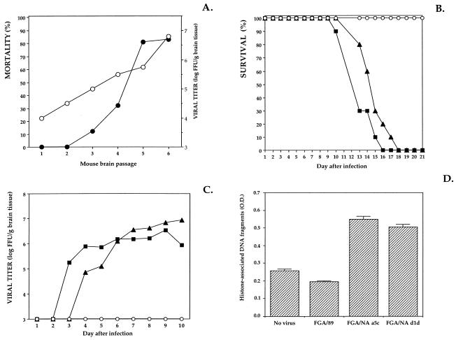
FIG. 1.
Replication of DEN virus in mouse brain. Litters of 2-day-old Swiss mice (Breeding Centre R. Janvier, Le Genest St-Isle, France) were inoculated i.c. with 20 μl of DEN virus in Leibovitz L-15 growth medium containing 2% heat-inactivated fetal bovine serum. (A) Mice were inoculated with 105 FFU of mouse-passaged DEN-1 virus (strain FGA/89) and observed daily for 21 days, and mortality was recorded (•). Eight days after inoculation, brains from three suckling mice were collected and weighed. Brain tissues were prepared as 10% (wt/vol) suspensions, and their infectivity was titrated by FIA (○). Titers are expressed as FFU per gram of brain tissue. (B and C) Newborn mice were inoculated with 5,000 FFU of FGA/89 (○), FGA/NA a5c (▪), or FGA/NA d1d (▴). For panel B, infected mice were observed daily for 21 days and mortality was recorded. For panel C, viral growth in the brains of infected mice was titrated. Each point represents the titration of pooled brain tissues extracted from three DEN virus-infected mice. (D) Oligonucleosomal DNA fragments in brain tissue suspensions were quantified by ELISA. Mouse brains were harvested in triplicate 9 days after infection. Brain tissue suspensions (20 mg) were incubated with lysis buffer, and the histone-associated DNA fragments released into the cytoplasmic fractions were quantified with a cell death detection ELISA kit according to the manufacturer’s protocol (Boehringer Mannheim Biochemicals). Optical density (O.D.) was measured at 405 nm.

