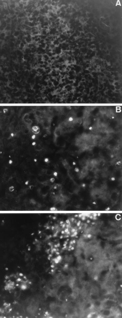
FIG. 2.
Distribution of apoptosis in the brain regions of DEN virus-infected mice. Mouse brains were harvested 9 days after inoculation. For mouse brain sections, the brains were removed, covered in Tissue-Tek O.C.T. (Miles) embedding medium, and stored at −80°C. Parasagittal sections of the brain blocks (15-μm thickness) were cut on a cryostat (Jung Frigocut) and mounted onto Vectabond-precoated slides (Vector Laboratories). Sections were fixed in 3% paraformaldehyde in phosphate-buffered saline and stored at 4°C in 70% ethanol. Tissue sections mock infected (A) or infected with DEN virus (FGA/NA d1d) (B and C) were processed for TUNEL analysis. The TUNEL assay was performed with mouse brain sections as described in the instructions to an in situ cell death detection kit from Boehringer Mannheim Biochemicals. TUNEL-positive cells were observed by fluorescence. Cortical (A and B) and hippocampal (C) regions are shown. Magnification, ca. ×100.

