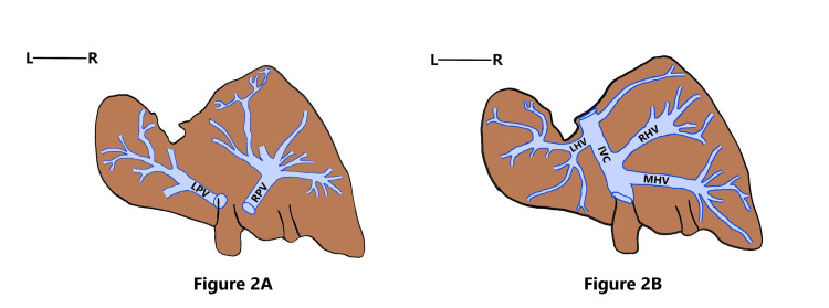
Figure 2. Schematic diagram showing the venous pattern of Riedel’s lobe and the beaver tail variant.
Figure 2A: Schematic diagram depicting the portal venous radicals supplying Riedel’s lobe and the beaver tail variant. Figure 2B: Schematic diagram depicting the hepatic venous radicals draining Riedel’s lobe and the beaver tail variant
RPV: right branch of the portal vein; LPV: left branch of the portal vein, RHV: right hepatic vein; MHV: middle hepatic vein; LHV: left hepatic vein; IVC: inferior vena cava

