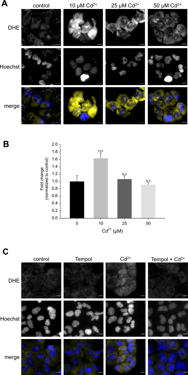
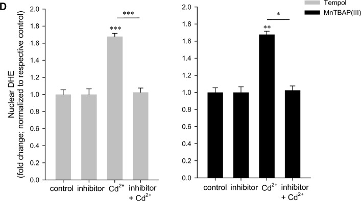
Fig. 3.
Superoxide anions predominate at low Cd2+. WKPT-0293 Cl.2 cells were exposed to Cd2+ for 1 h and superoxide anions were subsequently detected by dihydroethidium (DHE), microscope analysis (A) (scale bar = 20 µm) and quantification of thresholded images (B) (n = 5). C Pre-treatment with Tempol abolished superoxide anion generation by 10 µM Cd2+. Scale bar = 20 µm. Images are representative of 5 independent experiments. D Quantification of DHE oxidation in the nucleus with Tempol (n = 5) or MnTBAP (n = 9). Statistical analyses using one-way ANOVA with Holm–Sidak posthoc test compares Cd2+ treated to control cells (B) or antioxidant+Cd2+ to Cd2+ only cells (D)


