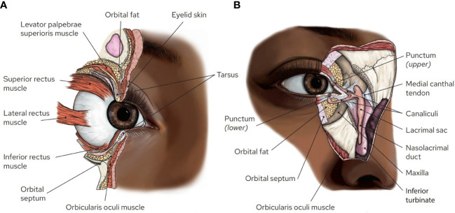
Figure 1.
Orbital and ocular adnexal anatomy. (A) Sagittal section of the orbit illustrating the proximity of multiple structures within the eyelids and anterior orbit. Note the orbital septum, which separates the preseptal tissues from the orbital tissues, and the presence of fat that cushions the orbital contents prevents establishing clear margins within the orbit. (B) En face view of the medial orbit with portions of the skin, muscle, orbital septum, medial canthal tendon, and bone cut away to illustrate the lacrimal drainage system. Artwork by Rae Senarighi.

