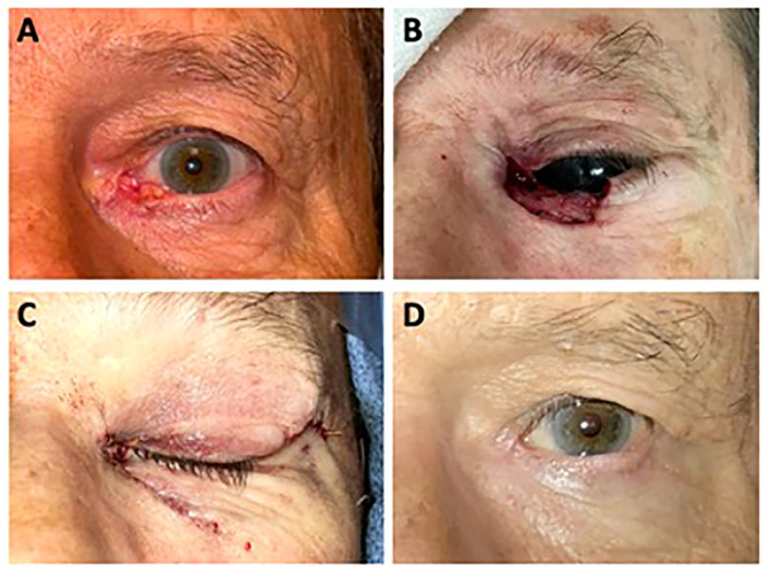
Figure 2.
(A) A 69-year-old man presented with a non-healing, ulcerated left lower lid lesion with raised, pearly, telangiectatic borders. Biopsy revealed basal cell carcinoma. (B) Surgical defect of Mohs micrographic surgery. (C) Immediately following surgical reconstruction of the left lower lid margin using a semicircular Tenzel flap and reconstruction of the left upper lid margin with a medial tarsal strip. (D) 12 months following Mohs surgery and reconstruction.

