. 2024 Jan 9;12:RP90136. doi: 10.7554/eLife.90136

Figure 4. Bsh with Notch signaling activates Ap and specifies L4 neuronal fate.

(A–D) Ectopic expression of N-ICD in newborn L5 neurons (Bsh-Gal4 >UAS-N-ICD) results in ectopic Hey and Ap activation and an increased number of L4 neurons at 19 hr APF. Here and below, scale bar, 10 µm, n≥5 brains. (E–J) N-ICD shows Bsh+ lamina neurons are mainly Ap+ L4 neurons, though the number of Bsh+ lamina neurons remains unaffected at 3-4d APF. (I–J) Quantification (single optical section). (K) Summary. Data are presented as mean ± SEM. Each dot represents each brain. n=6 brains in (I), (J). ***p<0.001, ns = not significant, unpaired t-test.

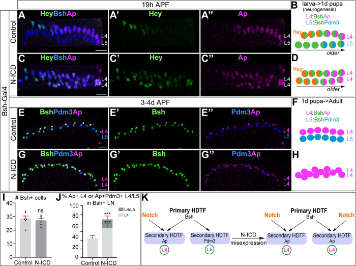
Figure 4.

Figure 4—figure supplement 1. Temporally restricted activation of Notch signaling by 27G05-Gal4 enables Bsh to specify L4 neuronal fate over L5.

Figure 4—figure supplement 1.

(A) Schematic of the genetics for temporally restricted activation of Notch signaling. (B-C”) Activation of Notch signaling from 0 hr APF (affected region circled) results in ectopic Hey expression at 19 hr APF. Here and below, scale bar, 10 µm, n≥5 brains. (D–G) Activation of Notch signaling from 0 hr APF (affected region circled) results in no change to Bsh expression, loss of Pdm3+ L5 neurons, and gain of Ap+Pdm3 L4 neurons at 3-4d APF. (F and G) Quantification (single optical section). Data are presented as mean ± SEM. Each dot represents each brain. ***p<0.001, ns = not significant, unpaired t-test.
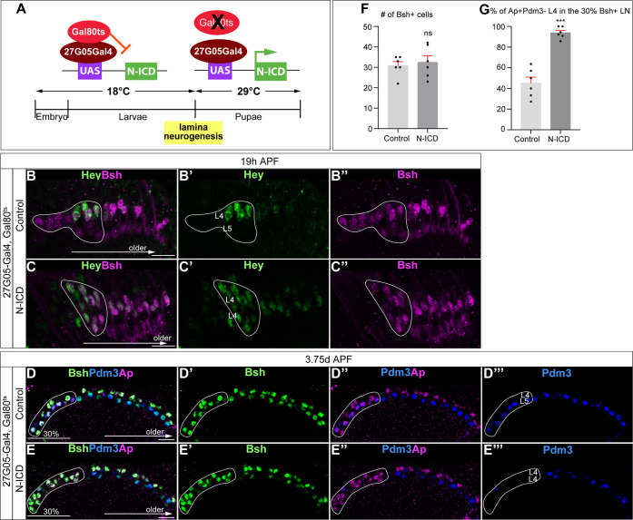
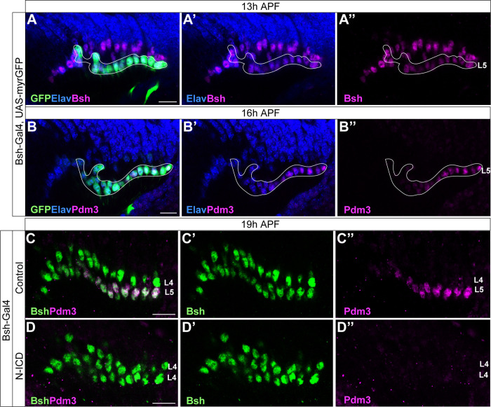
Figure 4—figure supplement 2. Bsh-Gal4 is expressed in newborn L5 neurons; Bsh does not activate L5 marker Pdm3 when Notch signaling is activated in newborn L5 by Bsh-Gal4 and UAS-N-ICD.

Figure 4—figure supplement 2.

(A-B”) Bsh-Gal4 is expressed in newborn L5 neurons. (A-A”) GFP+ cells (white line circle; Bsh-Gal4 >UAS-myrGFP) label L5 neurons which are in the bottom (proximal) row of Bsh+ cells during lamina neurogenesis (13 hr APF). Here and below, scale bar, 10 µm, n≥5 brains. (B-B”) GFP+ cells (white line circle; Bsh-Gal4 >UAS-myrGFP) label Pdm3+ newborn L5 and early-born L5 neurons during lamina neurogenesis (16 hr APF). (C-D”) Bsh does not activate L5 marker Pdm3 when Notch signaling is activated in newborn L5 by Bsh-Gal4 and UAS-N-ICD. Pdm3 expression is initiated in L5 neurons in control but not in N-ICD (Bsh-Gal4 >UAS-N-ICD).
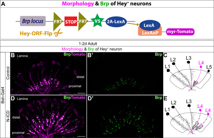
Figure 4—figure supplement 3. Bsh-Gal4 activation of Notch signaling in newborn L5 neurons specifies L4-like morphology and presynaptic sites.

Figure 4—figure supplement 3.

(A) Schematic of the genetics used to trace the morphology and presynaptic site (Brp) of Hey+ neurons. (B–E) In controls, Tomato+ neurons in lamina have L4 morphology and presynaptic sites labeled by Brp-V5. In Bsh-KD, Tomato+ cells, which trace the endogenous and ectopic Hey+ neurons, have L4-like morphology and presynaptic sites labeled by Brp-V5 in the lamina. Scale bar, 10 µm. n≥5 brains.