. 2024 Jan 11;12:RP87607. doi: 10.7554/eLife.87607

Figure 12. Uniform Manifold Approximation and Projection (UMAP) cluster analyses of cells from the hepatocellular carcinoma (HCC) and benign liver based on gene expressions with different standard deviation cutoffs.

(A) UMAP clustering of cells based on gene expression with standard deviations at least 0.5, 0.8, 1.0, or 1.4. The numbers of genes employed in the UMAP analysis are indicated. Blue dot, cell from the benign liver; red dot, cell from HCC. (B) UMAP clustering of cells based on isoform expressions with standard deviations at least 0.5, 0.8, 1.0, or 1.4. The numbers of genes employed in the UMAP analysis are indicated. Blue dot, cell from the benign liver; red dot, cell from HCC.

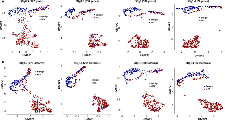
Figure 12.

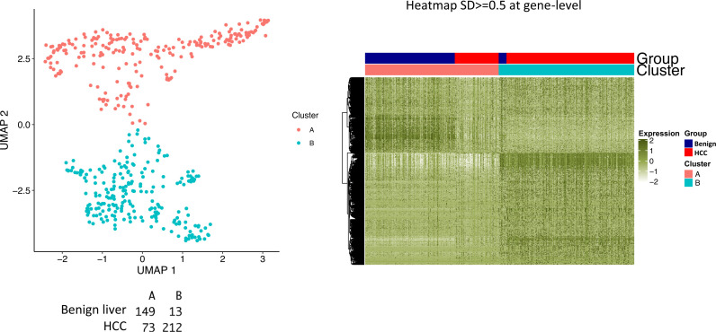
Figure 12—figure supplement 1. Segregation of cells between hepatocellular carcinoma (HCC) and benign liver samples.

Figure 12—figure supplement 1.

Relabeling of clusters as ‘A’ and ‘B’ based on gene expressions with a standard deviation ≥ 0.5. Left panel: Uniform Manifold Approximation and Projection (UMAP) clustering. The numbers of cells from the HCC and benign liver samples in each cluster are indicated. Right panel: heatmap of clusters A and B. Cells from HCC and benign liver are indicated.
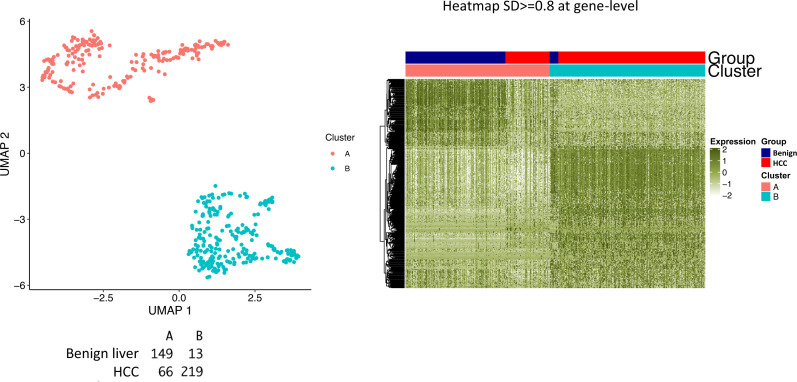
Figure 12—figure supplement 2. Segregation of cells between hepatocellular carcinoma (HCC) and benign liver samples.

Figure 12—figure supplement 2.

Relabeling of clusters as ‘A’ and ‘B’ based on gene expressions with a standard deviation ≥ 0.8. Left panel: Uniform Manifold Approximation and Projection (UMAP) clustering. The numbers of cells from HCC and benign liver in each cluster are indicated. Right panel: heatmap of clusters A and B. Cells from HCC and benign liver are indicated.
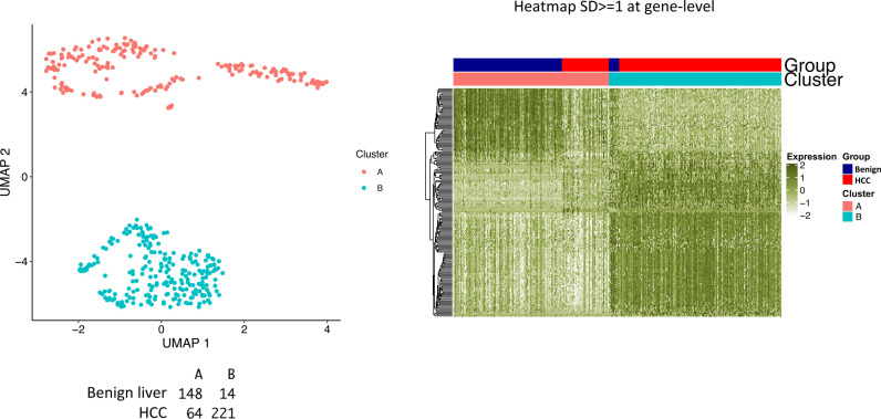
Figure 12—figure supplement 3. Segregation of cells between hepatocellular carcinoma (HCC) and benign liver samples.

Figure 12—figure supplement 3.

Relabeling of clusters as ‘A’ and ‘B’ based on gene expressions with a standard deviation ≥ 1.0. Left panel: Uniform Manifold Approximation and Projection (UMAP) clustering. The numbers of cells from HCC and benign liver in each cluster are indicated. Right panel: heatmap of clusters A and B. Cells from HCC and benign liver are indicated.
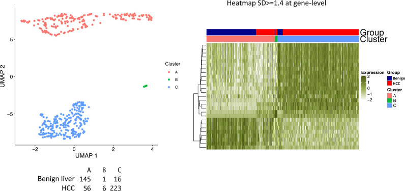
Figure 12—figure supplement 4. Segregation of cells between hepatocellular carcinoma (HCC) and benign liver samples.

Figure 12—figure supplement 4.

Relabeling of clusters as ‘A’, ‘B’, and ‘C’ based on gene expressions with a standard deviation ≥ 1.4. Left panel: Uniform Manifold Approximation and Projection (UMAP) clustering. The numbers of cells from HCC and benign liver in each cluster are indicated. Right panel: heatmap of clusters A, B, and C. Cells from HCC and benign liver are indicated.
Figure 12—figure supplement 5. Segregation of cells between hepatocellular carcinoma (HCC) and benign liver samples.

Figure 12—figure supplement 5.

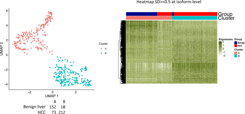
Relabeling of clusters as ‘A’ and ‘B’ based on isoform expressions with a standard deviation ≥ 0.5. Left panel: Uniform Manifold Approximation and Projection (UMAP) clustering. The numbers of cells from HCC and benign liver in each cluster are indicated. Right panel: heatmap of clusters A and B. Cells from HCC and benign liver are indicated.
Figure 12—figure supplement 6. Segregation of cells between hepatocellular carcinoma (HCC) and benign liver samples.

Figure 12—figure supplement 6.

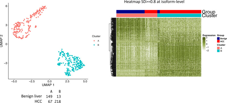
Relabeling of clusters as ‘A’ and ‘B’ based on isoform expressions with a standard deviation ≥ 0.8. Left panel: Uniform Manifold Approximation and Projection (UMAP) clustering. The numbers of cells from HCC and benign liver in each cluster are indicated. Right panel: heatmap of clusters A and B. Cells from HCC and benign liver are indicated.
Figure 12—figure supplement 7. Segregation of cells between hepatocellular carcinoma (HCC) and benign liver samples.

Figure 12—figure supplement 7.

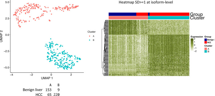
Relabeling of clusters as ‘A’ and ‘B’ based on isoform expressions with a standard deviation ≥ 1.0. Left panel: Uniform Manifold Approximation and Projection (UMAP) clustering. The numbers of cells from HCC and benign liver in each cluster are indicated. Right panel: heatmap of clusters A and B. Cells from HCC and benign liver are indicated.
Figure 12—figure supplement 8. Segregation of cells between hepatocellular carcinoma (HCC) and benign liver samples.

Figure 12—figure supplement 8.

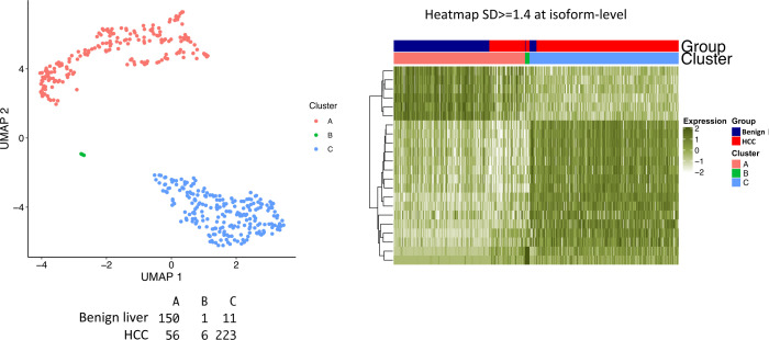
Relabeling of clusters as ‘A’, ‘B’, and ‘C’ based on isoform expressions with a standard deviation ≥ 1.4. Left panel: Uniform Manifold Approximation and Projection (UMAP) clustering. The numbers of cells from HCC and benign liver in each cluster are indicated. Right panel: Heatmap of clusters A, B, and C. Cells from HCC and benign liver are indicated.