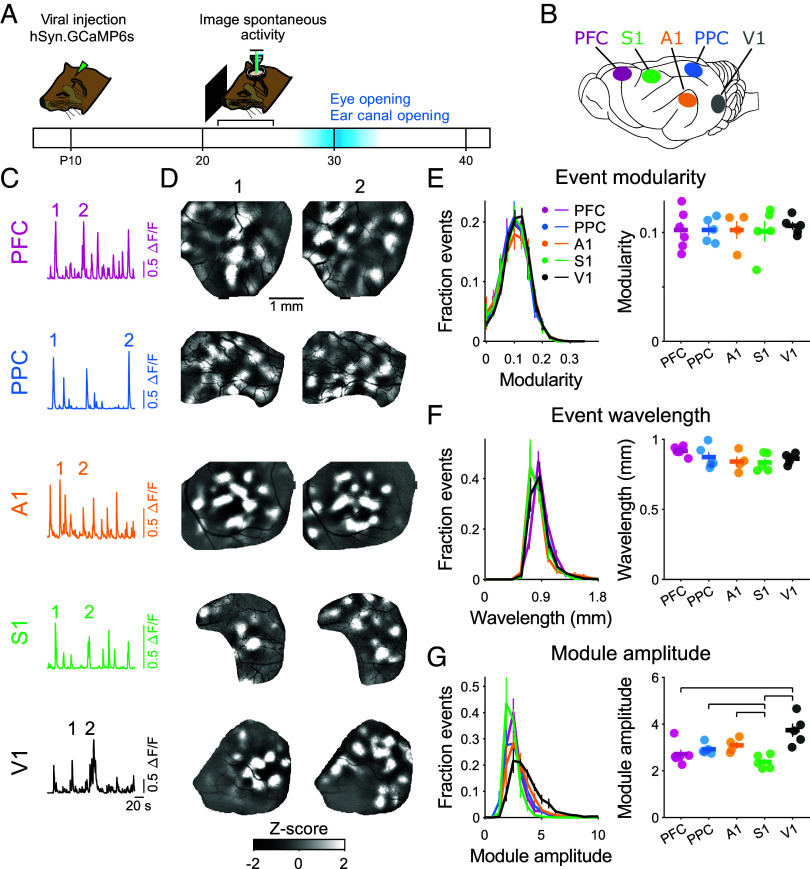
Fig. 1.
Spontaneous activity is highly modular in early development across diverse cortical areas. (A) Experimental schematic. Spontaneous activity was imaged at P21-24, 7 to 14 d prior to eye-opening and ear canal opening. (B) Activity was imaged in primary somatosensory (S1), auditory (A1), and visual (V1) cortices, and in the association areas PFC and PPC. (C) Time course of spontaneous activity (mean activity across ROI) in each brain area imaged in independent experiments. (D) Individual spontaneous events (times indicated in C) show highly modular activity in all areas. (E) The modularity of spontaneous events does not vary across cortical areas. For panels (E–G): Left plot shows distribution across all events, Right plot shows median of distribution for each animal (dots) and mean across animals (horizontal bar). (F) The wavelength of activity for spontaneous events is similar across events from different areas. (G) Module amplitude (active module vs. adjacent cortex) is generally similar across areas, with significantly lower amplitude in S1 and higher amplitude in V1. Significant post hoc pairwise comparisons indicated by horizontal lines. Error bars ± SEM.

