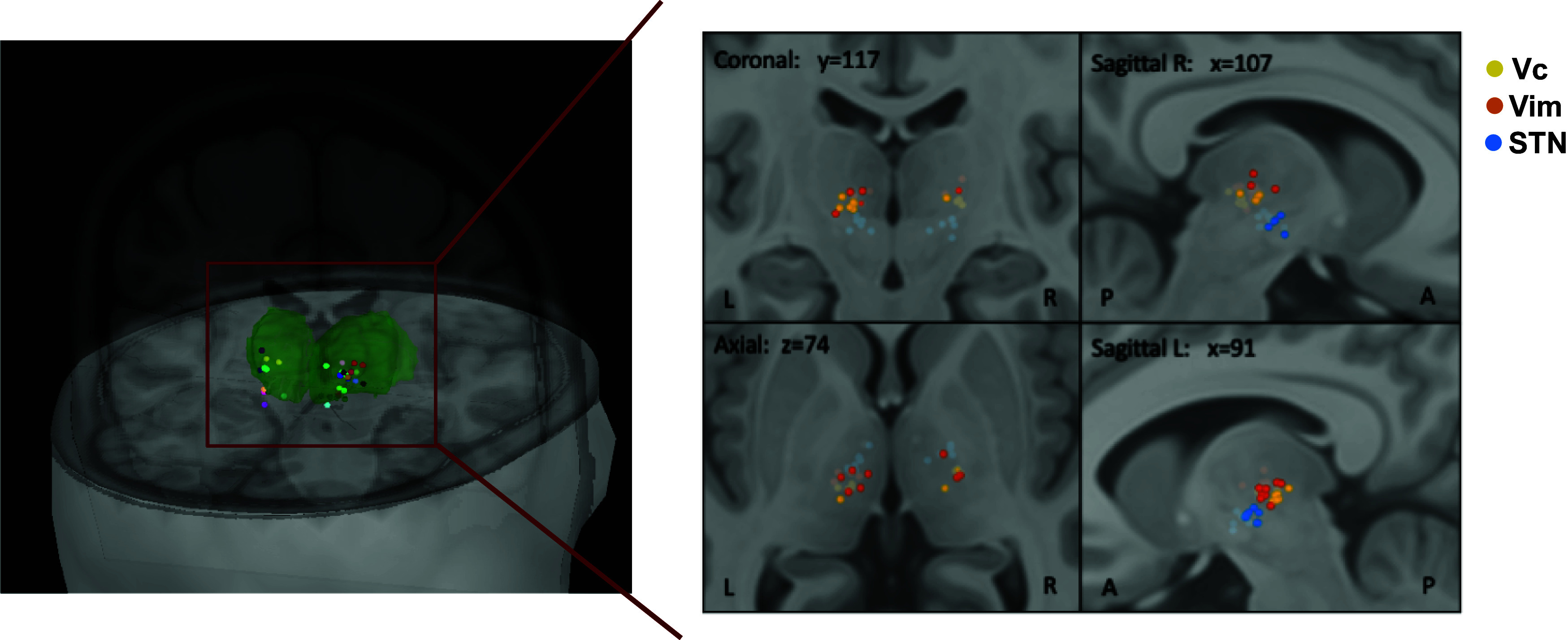
Fig. 1.

Recording sites. Location of recording electrodes across the 23 surgeries color-coded by patient (Left) and color-coded by identified region for Vc (yellow), Vim (orange), and STN (blue) (Right). Green volume represents the thalamus. Labeling of recording sites was obtained intraoperatively based on each patient’s brain scan, coordinates were then converted to MNI space for visualization and are projected here on the MNI/ICBM152 human brain template (69). Note that for some sessions, location dots overlap as different recordings were performed in the same location. Abbreviations used: ventral caudal thalamic nucleus (Vc), ventral intermedius thalamic nucleus (Vim), subthalamic nucleus (STN). Left and Right panels generated using Brainstorm (70).
