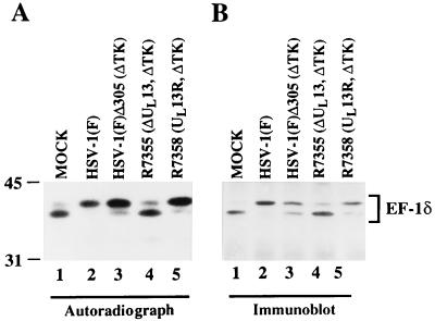
FIG. 5.
Autoradiographic and photographic images of 32P-radiolabeled infected-cell lysate immunoprecipitated by the antibody to EF-1δ, subjected to autoradiography, and then reacted with antibody to EF-1δ. Vero cells were mock infected or infected with the indicated virus. At 7 h after infection, the cells were labeled with 32Pi for 5 h and then harvested, solubilized, immunoprecipitated with the antibody to EF-1δ, electrophoretically separated in an SDS–9% polyacrylamide gel, transferred to a nitrocellulose sheet, and subjected to autoradiography (A) and then reacted with the antibody to EF-1δ (B). ΔTK, thymidine kinase gene deleted; ΔUL13, UL13 gene deleted; UL13R, UL13 gene repaired.

