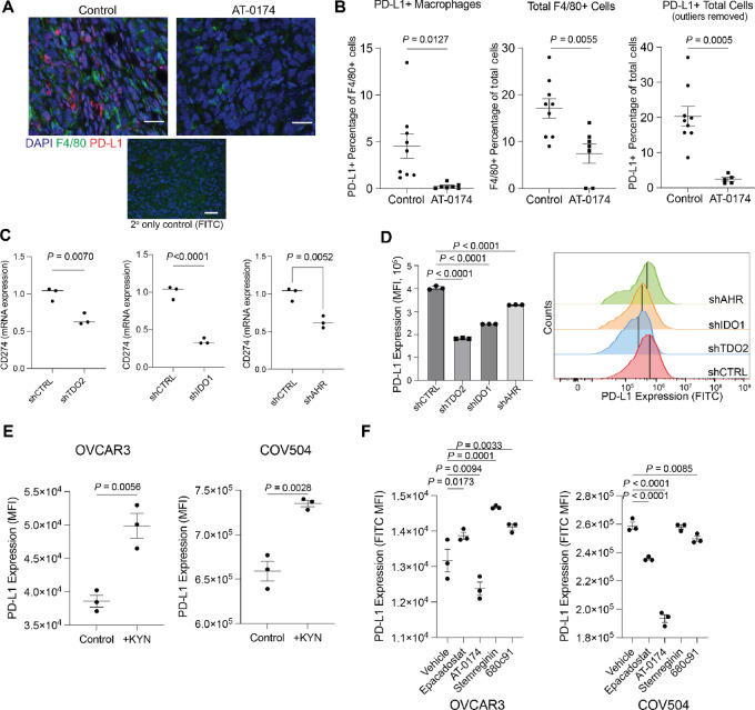
FIGURE 5.
A dual IDO/TDO2 inhibitor decreases PD-L1 expression. A, mIHC of ID8 tumors (described in Fig. 4) for F4/80 and PD-L1. B, Quantification of F4/80 and/or PD-L1+ cells. C,CD274 (PD-L1) expression in OVCAR3 following knockdown of TDO2, IDO1, AhR. Internal controls, 18S. D, PD-L1 protein expression measured via flow cytometry following knockdown of TDO2, IDO1, AhR in OVCAR3. E, PD-L1 protein expression measured via flow cytometry following exogenous supplementation of KYN (10 µmol/L) in OVCAR3 and COV504 cells. F, PD-L1 expression in TDO2 or IDO inhibitor epacadostat (10 µmol/L), AT-0174 (5 µmol/L), StemRegenin (10 µmol/L) and 680c91 (10 µmol/L) treated OVCAR3 and COV504 cells. Error bars, SEM. Statistical test, unpaired t test.

