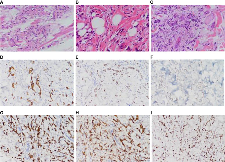
Figure 3.
Pathological picture. (A) Pathological images of the patient’s remnant stomach, partial jejunum, and partial resection specimen taken on April 30, 2020, (B) pathological images of the back skin lump taken on October 25, 2021, (C) pathological images of the left abdominal skin tissue taken on July 20, 2022, (D) immunohistochemical CK7 pathological images of the left abdominal skin tissue taken on July 20, 2022, (E) immunohistochemical CDX-2 pathological images of the left abdominal skin tissue taken on July 20, 2022, (F) Immunohistochemical STAB2 pathological image of left abdominal skin tissue taken on July 20, 2022, (G) Immunohistochemical CK8-18 pathological image of left abdominal skin tissue taken on July 20, 2022, (H) Immunohistochemical Villin pathological image of left abdominal skin tissue taken on July 20, 2022, (I) Immunohistochemical ki67 pathological image of left abdominal skin tissue taken on July 20, 2022.

