
FIGURE 1.
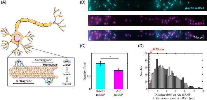
β‐actin and Arc mRNPs are packaged separately in dendrites. (A) Schematic of the motor‐driven transport of mRNP complexes in a neuron. An mRNP, in the form of a complex of mRNA bound to RBPs, is transported along microtubules by dynein and kinesin motor proteins. (B) Representative FISH images of β‐actin mRNA (cyan) and Arc mRNA (magenta) in the proximal dendrite. (C) Bar graph comparing the density of β‐actin and Arc mRNP per unit length (μm). In proximal dendrites, the density of β‐actin mRNP was significantly higher than that of Arc mRNP (* p < 0.05, pairwise t test; n = 54 dendrites). The error bars represent the standard error of mean (SEM). (D) Histogram of distances from an Arc mRNP to the nearest β‐actin mRNP (n = 1443 Arc mRNPs). The percentage of Arc mRNPs colocalized with β‐actin mRNP (within the threshold of 0.25 μm) was 0.14%.