
FIGURE 3.
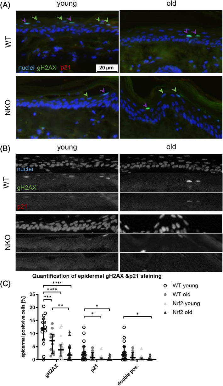
Assessment of activated DNA damage repair in epidermal cells positive for senescence marker p21. (A) Representative images of murine tail cross sections of young and old wildtype (WT) and Nrf2 deficient (NKO) animals. Shown are immunofluorescence stainings of the activated DNA damage repair marker gamma H2AX (gH2AX, green), the senescence marker p21 (red) and nuclei counterstain with Höchst reagent. Green arrow heads indicate gH2AX single positive and purple arrow heads double positive cells respectively. Scale bar: 20 μm. (B) Matrix of single channel greyscale image sections of the merged micrographs shown in A. (C) Plot of gH2AX and/ or p21 positive cells in the epidermis shown as percentage of total epidermal cells. Symbols represent quantification data from 4 field of view per mouse, 3 mice per genotype & age group. Asterisks indicate statistically significant differences (*p < 0.05, **p < 0.01, ***p < 0.001, ****p < 0.0001, one‐way ANOVA with Tukey's correction for multiple comparisons. Results are depicted as median with 95% confidence interval.)