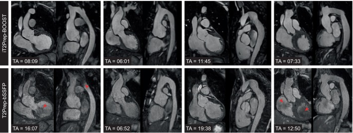
FIGURE 2.

Visual comparison between bright‐blood images produced by the proposed iT2Prep‐BOOST approach compared to the conventional T2prep‐bSSFP for four representative patients (columns) showing a coronal view and a reformatted long axis view of the thoracic aorta. Acquisition time (TA) is expressed in minutes:seconds. (a) A 35‐year‐old patient with bicuspid aortic valve and coarctation of the aorta. Flow artefacts at the level of the arch, the isthmus, and the left ventricular outflow tract (red arrows) are reduced; (b) 19‐year‐old patient with aortic root aneurysm. Respiratory artefacts in the aortic root are reduced in the proposed approach; (c) 20‐year‐old patient with aortic aneurysm in the descending aorta, post coarctation repair with end‐to‐end anastomosis; (d) 43‐year‐old patient with tortuous thoracic aorta post coarctation repair. Blood pool inhomogeneities (red arrows) are attenuated.
