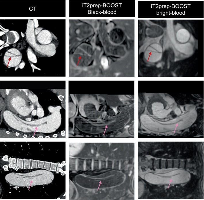
FIGURE 10.

Comparison between conventional CT and proposed iT2Prep‐BOOST MR imaging in 54‐year‐old patient with chronic aortic dissection. The dissection flap is complex and extends from the aneurysmal thoracic descending aorta to the left common iliac artery (pink arrow). It is demonstrated both in the proposed iT2prep‐BOOST and the CT images (red arrow). The false lumen is dilated and aneurysmal.
