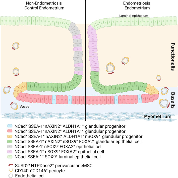
FIGURE 2.

Schematic showing the location of endometrial stem/progenitor cells in the basalis, functionalis and luminal epithelium of human endometrium based on specific surface markers of these cells demonstrating adult stem cell activity. Note the horizontal gland structure in the basalis gland from which emanates the vertical gland in the functionalis. It is likely that the NCAD−SSEA‐1+nSOX9+basalis epithelial cells re‐epithelialize the raw endometrial surface during menstruation to become the luminal epithelial cells. In endometriosis, most functionalis glandular epithelial cells are SSEA‐1+nSOX9+ (purple, right hand side), in contrast to normal endometrium, where they are only occasionally found in the functionalis (purple, left hand side). CD140b, PDGFRβ; eMSC, endometrial mesenchymal stem cells; NCAD, N‐cadherin; NTPDase2, nucleoside triphosphate diphosphohydrolase‐2 Source: Adapted from Salamonsen et al. 42 and Cousins et al. 43
