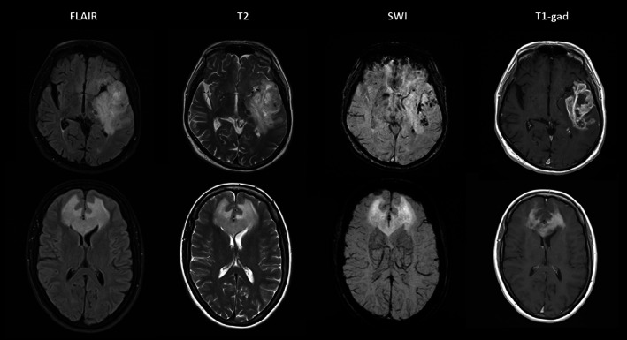
FIGURE 5.

Top row: 74‐year‐old man presenting with aphasia was found to have a high‐grade left temporal glioma on MRI (1.5 T). Note the intratumoral susceptibility signals (ITSS) on SWI indicative of microhemorrhage and vessel proliferation. Bottom row: 57‐year‐old woman presenting with behavioral changes due to a lymphoma. SWI does not show any ITSS despite marked homogeneous enhancement. 62
