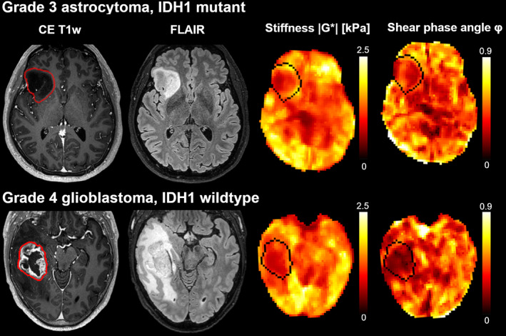
FIGURE 7.

Stiffness heterogeneity of gliomas. Contrast‐enhanced T1‐weighted images, FLAIR images, |G*| (stiffness), and φ maps (phase angle, related to viscosity) for two patients with gliomas. The images in the upper row are derived from a 40‐year‐old man with an IDH1‐mutated grade 3 astrocytoma, and the images in the lower row are derived from a 55‐year‐old man with an IDH1‐wild‐type grade 4 glioblastoma.
