FIGURE 9.

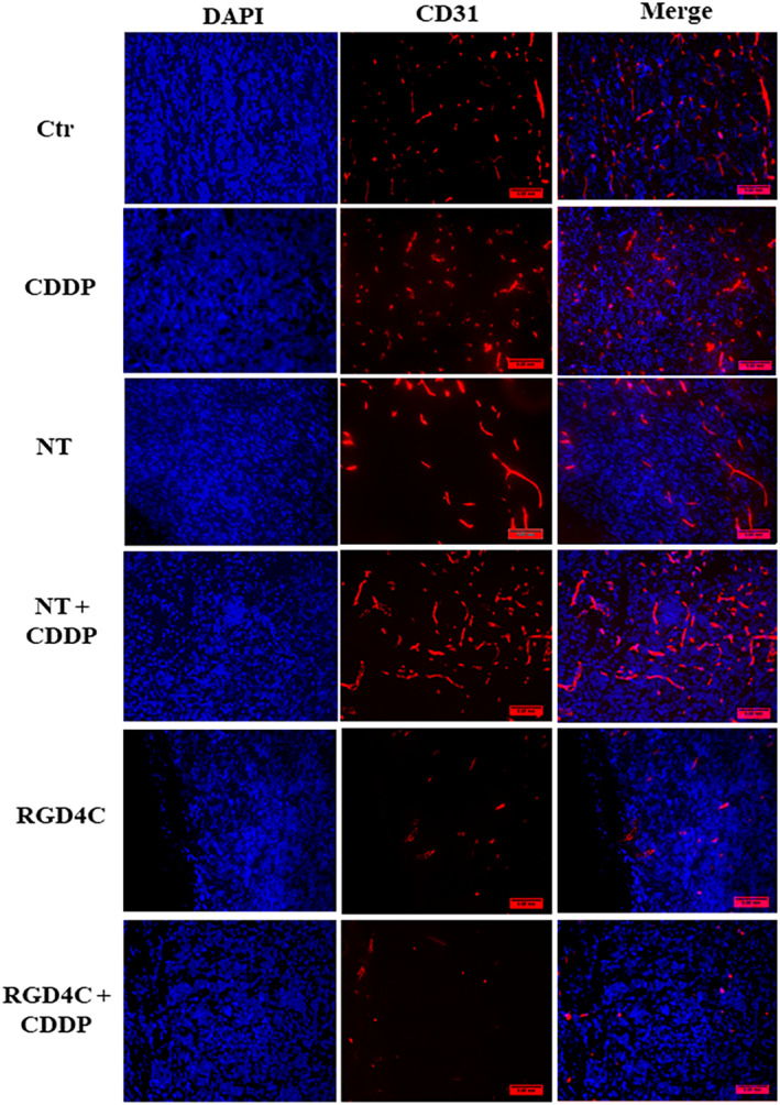
Immunohistochemistry analysis of the tumor vasculature. Tumor sections were stained with an anti‐mouse CD31 (red) as a blood vessel marker, and DAPI for nuclei staining (blue). Scale bar, 50 μm.

Immunohistochemistry analysis of the tumor vasculature. Tumor sections were stained with an anti‐mouse CD31 (red) as a blood vessel marker, and DAPI for nuclei staining (blue). Scale bar, 50 μm.