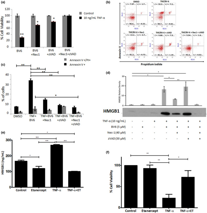
Figure 1.

A combination of TNF‐α and BV‐6‐induced HMGB1 release in HaCaTs which is modulated by inhibitors of both necroptosis (necrostatin) and apoptosis (Z‐VAD‐FMK). (a) HaCaT cell viability determined by MTT assay, (b) representative flow cytometric scatter plot of AV‐ and PI‐stained cells with (c) percentage cells positive stained and (d) extracellular HMGB1 following 24 h TNF‐α exposure ±40 μM necrostatin, 50 μM ZVAD (exemplar western blot image selected from n = 3). Densitometric analysis was performed on three separate blots and statistical analysis was undertaken using the Mann–Whitney U test (*P ˂ 0.05 and **P ˂ 0.01). (e) Supernatant HMGB1 levels determined by ELISA and (f) corresponding HaCaT viability by MTT assay (with BV‐6 pre‐treatment). Data represent mean normalized to untreated control (±SE) of three separate experiments conducted in triplicate (*P ˂ 0.05, **P ˂ 0.01, ***P < 0.001 and # < 0.05 vs. TNF‐a/BV‐6 treated cells).
