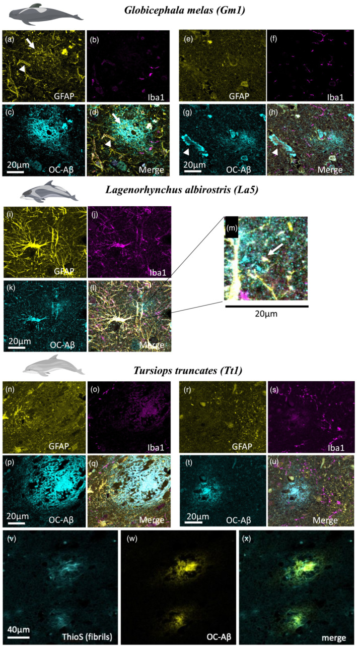
FIGURE 4.

GFAP (yellow), Iba1 (magenta) and Aβ fibril (cyan) immunofluorescence labelling in Globicephala melas (Gm1) (a–h), Lagenorhynchus albirostris (La5) (i–m) and Tursiops truncatus (Tt1) (n–u). Gm1 shows diffuse plaques (d,h), evidence of CAA (g, arrowhead) and localized presence of astrocyte processes near some (a, arrow) but not all (e) plaques. Microglia are located near to, but not within plaques (b,f). La5 shows extremely strong GFAP labelling (i), which bled through to other channels. Small diffuse plaques were present, including next to blood vessels (zoomed in section part label m, arrow) but no strong evidence of plaque‐associated gliosis. Tt1 showed more punctate astrocyte labelling (n,r) with little association with plaques despite the presence of many diffuse (u) and the occasional dense‐core (q) plaques. Although there was no association of microglia with the dense‐core plaque (o), there were some microglial processes near the edge of a diffuse plaque (s). We confirmed dense‐core plaques contained fibrils using Thioflavin S staining (v), which appeared in amyloid plaques stained for Aβ (w,x). Scale bars = 20 or 40 μm as indicated on panels
