
FIGURE 1.
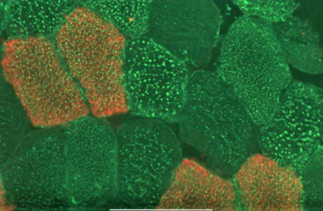
A representative light micrograph of a cryosection from a vastus lateralis muscle biopsy obtained from an IGR individual. After fixation in formalin, sections were washed 3 times in PBS and blocked using 10% goat serum. They were then incubated with anti‐hMYH7, followed by dye‐labeled secondary antibodies (red) to visualize the type I fibers. They were further stained with Bodipy 493/503 (green) in the dark. They were then washed in PBS, dried, covered with Prolong Gold™, and mounted. The bar indicates 100 μm.