
Figure 1. Adipose Tissue Organogenesis.
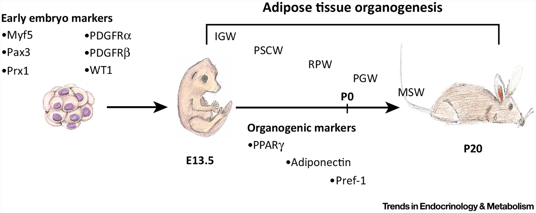
White adipose tissues are formed embryonically and postnatally. A variety of tools have been used to broadly identify early embryonic markers of adipose tissue, such as Myf5, Pax3, and Prx1. Around embryonic day (E)13.5, subcutaneous inguinal and periscapular adipose tissues are specified, patterned, and formed by more restrictive adipose lineage markers, such as PPARγ, adiponectin, and Pref-1. As embryogenesis continues (E18.5), the retroperitoneal adipose depot becomes specified, patterned, and formed. Both the perigonadal and mesenteric white adipose tissues are specified postnatally, beginning at postnatal day 2 (P)2 and ending near P20. Within the first month of life, all adipose depots are specified, formed, lipid filled, and functional. Designation of white adipose depots: IGW, Inguinal; MSW, mesenteric; PGW, perigonadal; PSCW, periscapular; RPW, retroperitoneal.