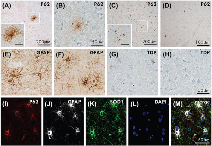
FIGURE 1.

p62‐positive astrocytes in CuATSM treatment group. p62‐immunoreactive astrocytes were observed in the motor cortex of ALS‐TDP (A: Case #3) and ALS‐SOD1 (B: Case #1) cases that had received CuATSM but not in ALS‐TDP (C: Case #11) or ALS‐SOD1 (D: Case #8) cases that had not received CuATSM treatment. The p62‐positive astrocytes were of similar appearance to GFAP‐positive astrocytic morphology (E: Case #3, F: Case #1). pTDP‐positive astrocytes were not seen in ALS‐TDP (G: Case #3) and ALS‐SOD1 (H: Case #1) cases. Immunofluorescent triple labelling demonstrated co‐localisation of p62, GFAP and SOD1 in the p62‐immunopositive astrocytes (I–M). A and C inset scale is 50 μm.
