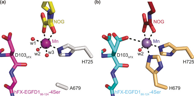
Figure 3.

A single protein‐bound ligand (i.e. H725) supports the metal ion in the H679A AspH variant. Colors: red: oxygen; blue: nitrogen. w: water. a) Active site view of the H679A AspH:Mn:NOG:hFX‐EGFD186‐124‐4Ser structure (colors: grey: H679A AspH; violet: Mn; yellow: carbon‐backbone of NOG; magenta: carbon‐backbone of the hFX‐EGFD186‐124‐4Ser peptide); b) active site view of the reported wt AspH:Mn:NOG:hFX‐EGFD186‐124‐4Ser structure (colours: bronze: wt AspH; lavender blue: Mn; maroon: carbon‐backbone of NOG; cyan: carbon‐backbone of the hFX‐EGFD186‐124‐4Ser peptide; PDB ID: 5JQY). [7]
