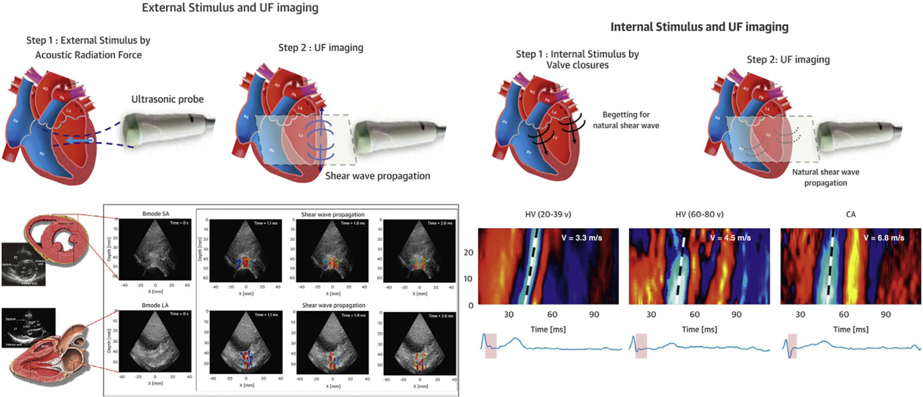
FIGURE 3. Shear-Wave Imaging.

(Left) SWI based on acoustic radiation force, which allows exact timing of the shear wave at end-systole or end-diastole. (Right) SWI of natural mechanical waves associated with cardiac mechanical events such as valve closure. CA = cardiac amyloidosis; HV = healthy volunteers; SWI = shear-wave imaging; UF = ultrafast. Adapted from Tamborini et al7 with permission.
