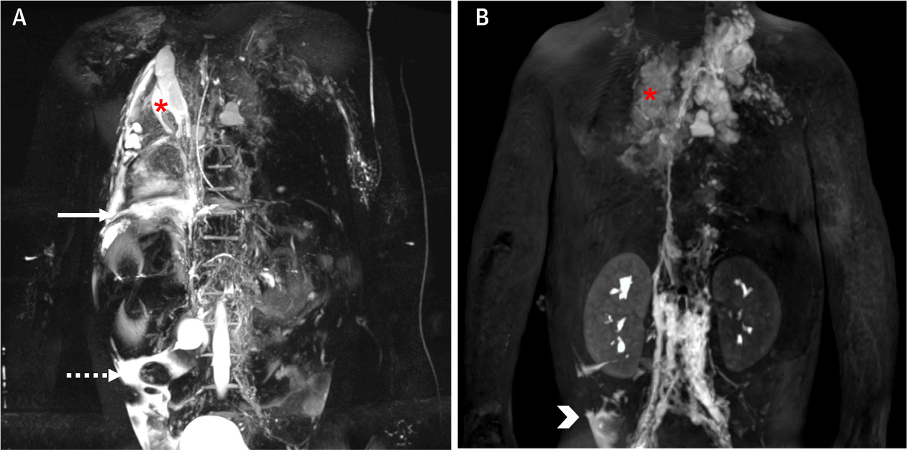
FIGURE 7. Magnetic Resonance Lymphangiography.

(A) 3D T2-weighted turbo spin echo acquisition and (B) still frame from a 3D T1-weighted sequence 14 minutes after injection during a dynamic contrast-enhanced magnetic resonance lymphangiography (DCMRL) acquisition in a 4-year-old patient with failing Fontan, plastic bronchitis, and chronic chylous effusions. The static T2-weighted images show severely abnormal thoracic lymphatics with right pulmonary lymphangiectasia (asterisk), as well as a right-side pleural effusion (solid arrow) and ascites (dashed arrow). The DCMRL shows retrograde lymphatic flow to both lungs, especially to the right (asterisk), as well as a subhepatic peritoneal lymphatic leak (arrowhead). Images provided by C. Lam, Toronto, Canada.
