
FIGURE 8. CCT Images With the Use of Low Radiation.
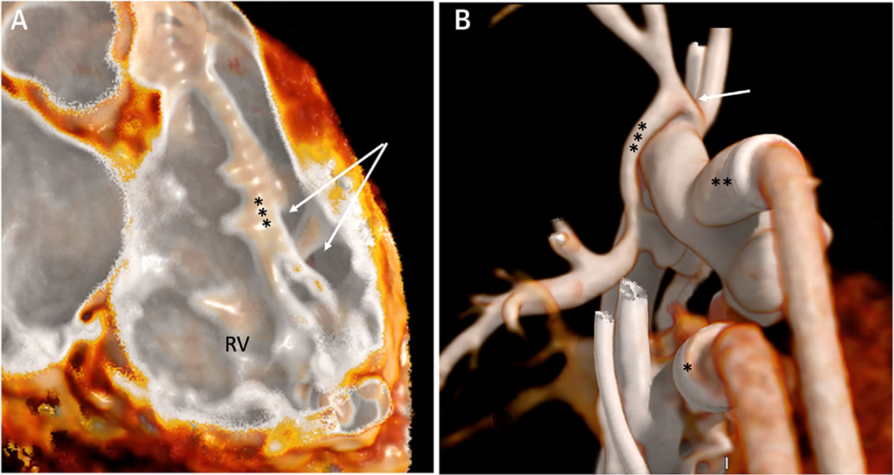
Electrocardiography-gated CCT images using single heart beat with low radiation. (A) Intraluminal view of the right ventricle (RV) showing a muscular ventricular septal defect (arrows) posterior to the septomarginal trabeculation (***). (B) External 3D reconstruction of venous (*) and arterial (**) ventricular assist device cannula in a patient with single-ventricle congenital heart disease. Narrowing of the innominate artery (arrow) is noted proximal to the systemic-to-pulmonary artery shunt (***) insertion. CCT = cardiac computed tomography.