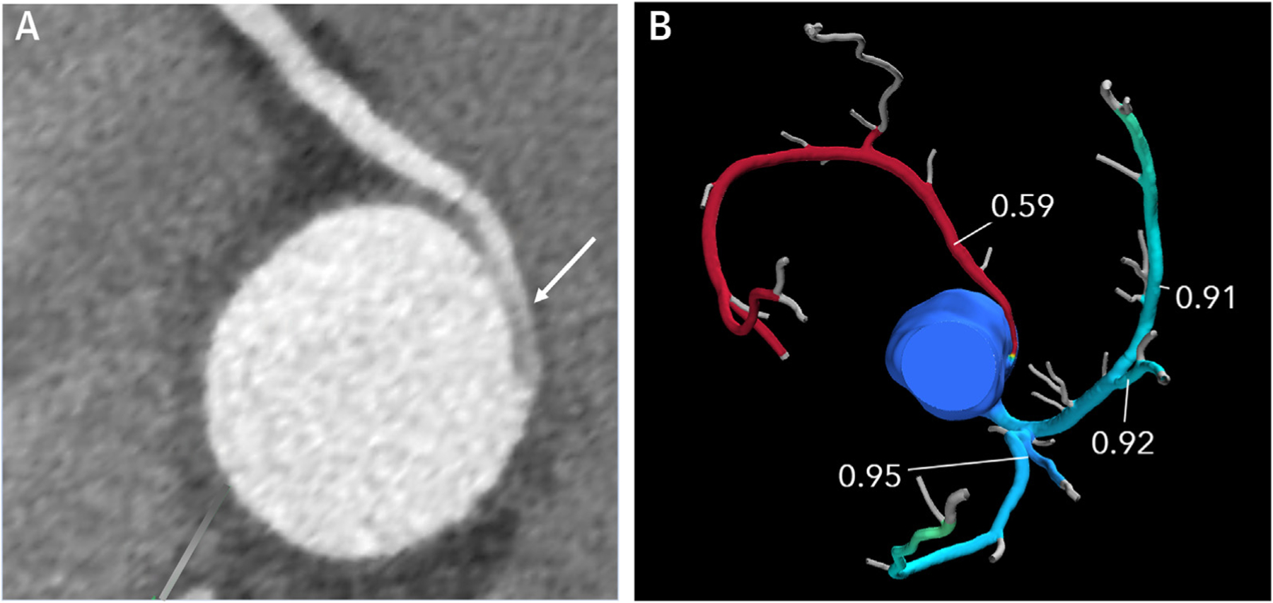
FIGURE 9. Computed Tomographic Fractional Flow Reserve (CT FFR).

(A) 2D short-axis CCT image of anomalous aortic origin of the right coronary artery from the ascending aorta just above the left sinus of Valsalva with an intramural proximal course (arrow) and normalization of vessel size as it leaves the aortic wall. The scan was performed using a dual source scanner with diastolic acquisition after administration of sublingual nitroglycerin. (B) CT FFR shows significant limitation to flow in the right coronary artery with a measurement of 0.59 at 2 cm distal to the anomalous origin.
