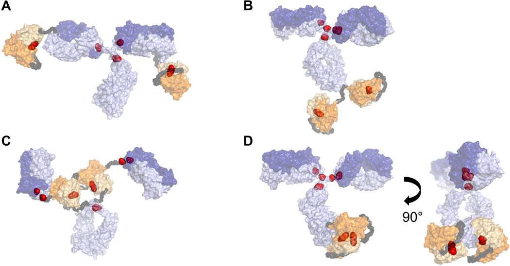
Figure 2.

Putative structural homology models for (a) BisAb-A, (b) BisAb-B, (c) BisAb-C, and (d) BisAb-D, which includes two orientations to observe the scFv fragments. Surface representations are shown for the full-length parental mAb (purple), scFv fragments (orange), and Gly/Ser linker peptides (dark gray). Interchain IgG and interdomain scFv disulfide molecules are represented as red spheres. Heavy chain and light chain sequences are depicted in light and dark shades, respectively. BisAb, bispecific antibody; mAb, monoclonal antibody; scFv, single-chain Fv
