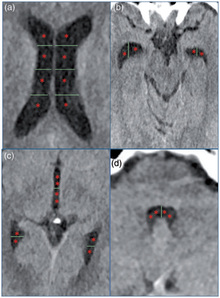
Figure 1.
Visual depiction of modified Graeb score.17 Images depict ventricular regions scored in the modified Graeb score. If a specific ventricular compartment is affected by hemorrhage, a point (one asterisk represents one point) is given. (a) Both right and left lateral ventricles are scored on a 4-point scale. (b) Right and left anterior temporal tips are scored on a 2-point system. (c) Posterior temporal tips are scored on a 2-point system, and the third ventricle is scored on a 4-point system. (d) The fourth ventricle is scored on a 4-point system. If the ventricle is completely filled with hemorrhage to the point where it is expanded, then an additional point is given for each ventricular region. The maximum score possible is 32 in which every region is filled with blood and expanded.

