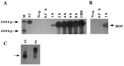
FIG. 2.
RNase protection and Northern blot analyses indicating the kinetics of IE3C transcription. (A) Autoradiogram from an RNase protection assay of cell lysates collected from uninfected cells (Neg.), from cycloheximide treated infected cells (CHX), and from infected cells at 0.5, 1, 2, 3, 4, 6, and 8 h p.i. (lanes 0.5 h through 8 h). Bands represent the 453-nt antisense IE3C-protected RNA fragment (2-day film exposure) (see Fig. 1). Lane M, molecular size markers; lane 3C, undigested probe. (B) IE3C detection at 0.5 and 1 h p.i. (7-day film exposure). (C) Northern blotting of poly(A) RNA from CCV-infected cells harvested at 3 and 8 h p.i., using the IE3C antisense riboprobe. The 1,400-nt band corresponding to the IE3C transcript is indicated by an arrow. The predominant banding at 8 h p.i. ranges from 2,000 to 3,500 nt.

