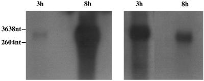
FIG. 8.
Northern blotting of poly(A) RNA from CCV-infected cells harvested at 3 and 8 h p.i., using the ORF39 riboprobe. (Left panel) Autoradiograph demonstrating expression levels at 3 and 8 h p.i. X-ray film was exposed for 2 h. (Right panel) Use of different film exposure times to demonstrate the size shift of the riboprobe-specific mRNA. The 3-h-p.i. portion was autoradiographed for 14 h, and the 8-h-p.i. portion was autoradiographed for 20 min. The positions of molecular size markers are shown on the left.

