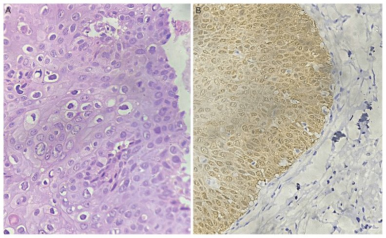
Fig. 6.
(A) Histopathological sections reveal acanthosis with loss of polarity, full thickness nuclear atypia, and increased mitotic activity, suggestive of VIN 3 (×400 magnification). (B) Immunohistochemistry reveals block positivity for p16 (×400 magnification). VIN, vulvar ntraeithelial neoplasia.

