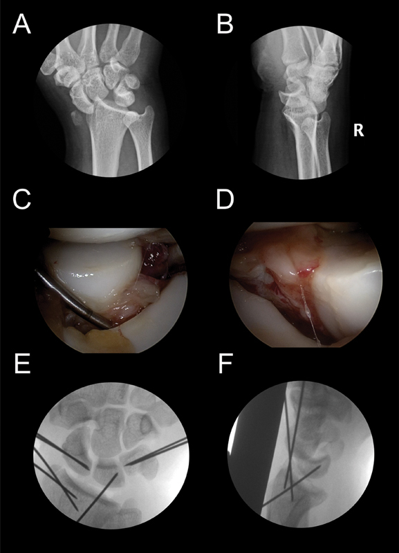
Fig. 21.

( A and B ) A transstyloid perilunate injury, X-ray view. ( C ) View from the MCU portal of the complete detachment of SL ligament. ( D ) View from the MCR portal of the complete detachment of LT ligament. ( E ) Two K-wires are introduced in the scaphoid up to the SL joint and another two in the triquetrum, again up the LT joint to but not crossing them. ( F ) If the lunate is too unstable, another K-wire is introduced from the radius to the lunate prior to the fixation of the LT or SL joints.
