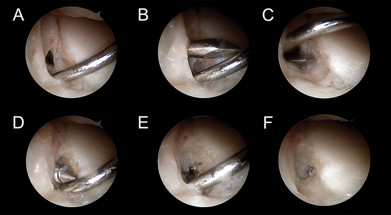
Fig. 6.

( A ) The arthroscope is in the MCU portal and a probe separates the soft tissues through the MCR portal for better visualization of the anchor insertion. ( B and C ) The hole for the anchor is made at the volar SL ligament insertion. ( D and F ) The anchor is introduced from the VR portal.
