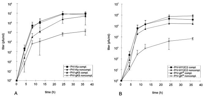
FIG. 6.
One-step growth analysis. Complementing and noncomplementing cells were infected at an MOI of 10 with PrV-Ka or PrV-gKβ (A) or at an MOI of 5 with PrV-9112C2 or PrV-gKgB (B) for 1 h at 4°C. After an additional 2 h at 37°C, remaining extracellular virus was inactivated by low-pH treatment. Immediately thereafter (0 h) and at the time points indicated, cells and supernatant were harvested, and progeny virus was titrated on complementing C53/54 cells. Mean values and standard deviations of results of two independent experiments are shown.

