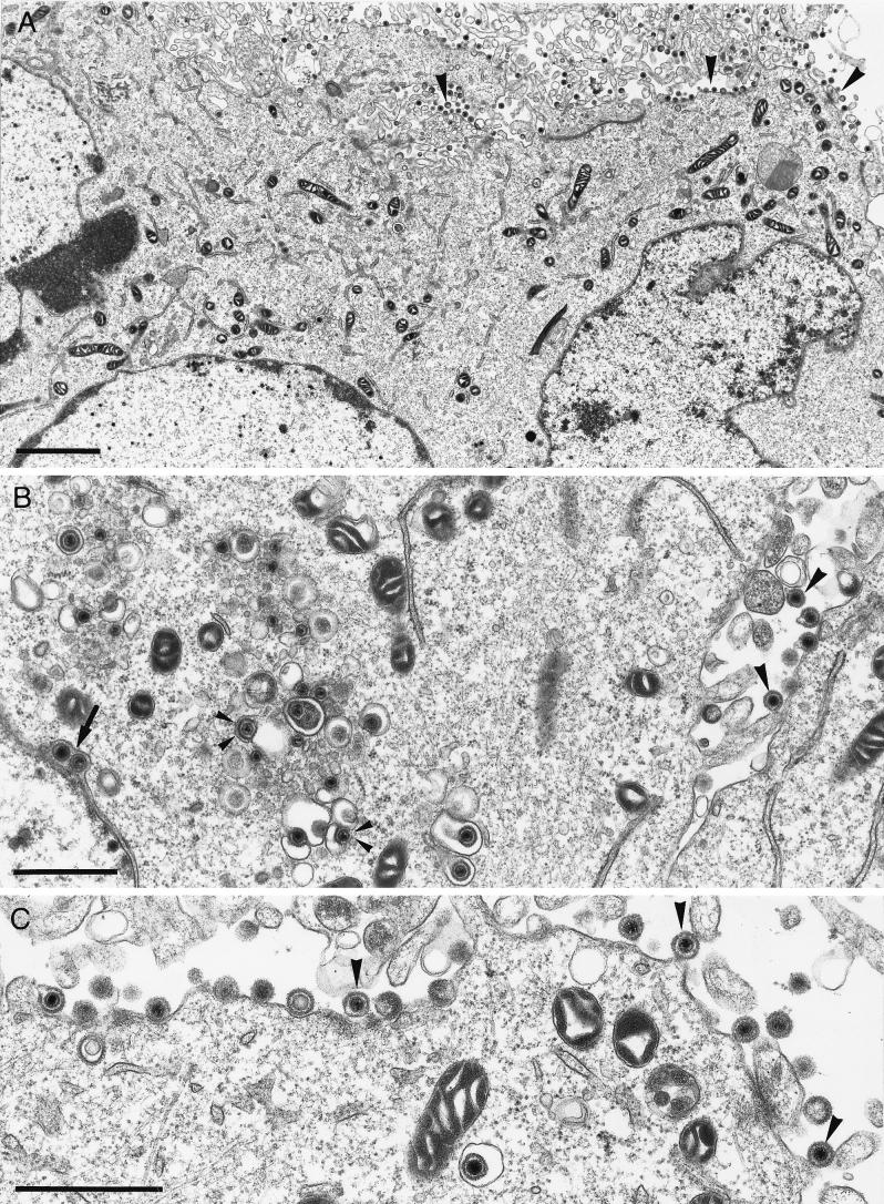
FIG. 7.
Electron microscopy of PrV-gKβ on complementing cells. B5′-64 cells were infected with PrV-gKβ at an MOI of 1 and analyzed 16 h p.i. The arrow shows primary envelopment in the perinuclear cisterna (B), small arrowheads indicate secondary envelopment in the Golgi area (B), and large arrowheads point to free virions in the extracellular space (A to C). Bars: A, 2.5 μm; B, 1 μm; C, 1 μm.

