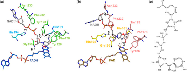
FIGURE 6.

Hydrogen bond interactions at the NAD+/H binding sites in the two homodimers of the complex NQO1‐NAD+/H. (a) Stick representation of the molecule NAD+/HD, the FADH− and all residues involved in the formation of the NAD+/H binding site of the homodimer C:D. (b) Stick representation of the molecule NAD+/HB, the FAD and all residues involved in the formation of the NAD+/H binding site of the homodimer A:B. The color code of the protein residues is the same as that shown in Figure 2. The color code of the FADH− and the FAD is the same as that shown in Figure S6. All hydrogen bond interactions established between the NAD+/H molecules and both the protein residues, and the FADs have been labeled and shown as black dashes. The pi‐interactions are labeled and dashed in red. (c) Two‐dimensional representation of the chemical structure of the NADH molecule.
