
Figure 2:
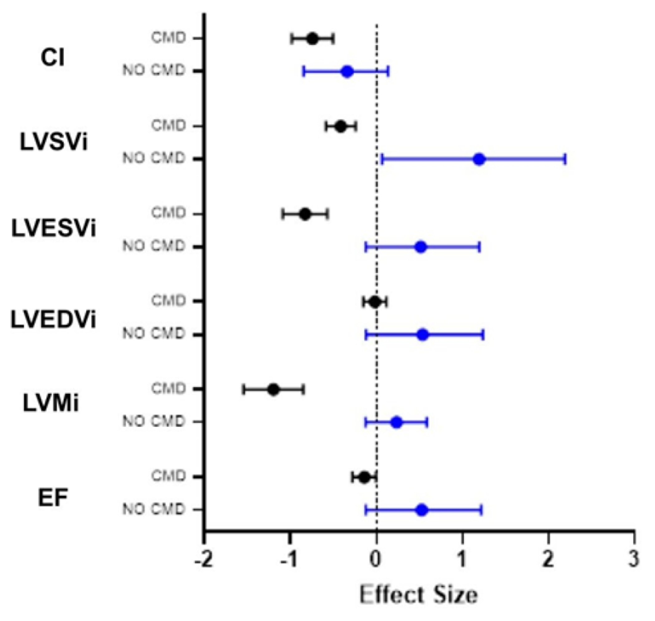
Patients without CMD (no CMD) show a stronger contribution to differences in cardiac structure and function between healthy controls and SLE patients.
Patients with no CMD (blue) on MPRI show a stronger contribution to overall effect of SLE on cardiac function than do patients with CMD (black bars) when compared with reference controls. Data is represented as Cohen’s d + C.I. CI: cardiac index; LVSVi: left ventricular systolic volume index; LVESVi: left ventricular end-systolic volume index; LVEDVi: left ventricular end-diastolic volume index; LVMi: left ventricular mass index; EF: ejection fraction.