Fig 2.
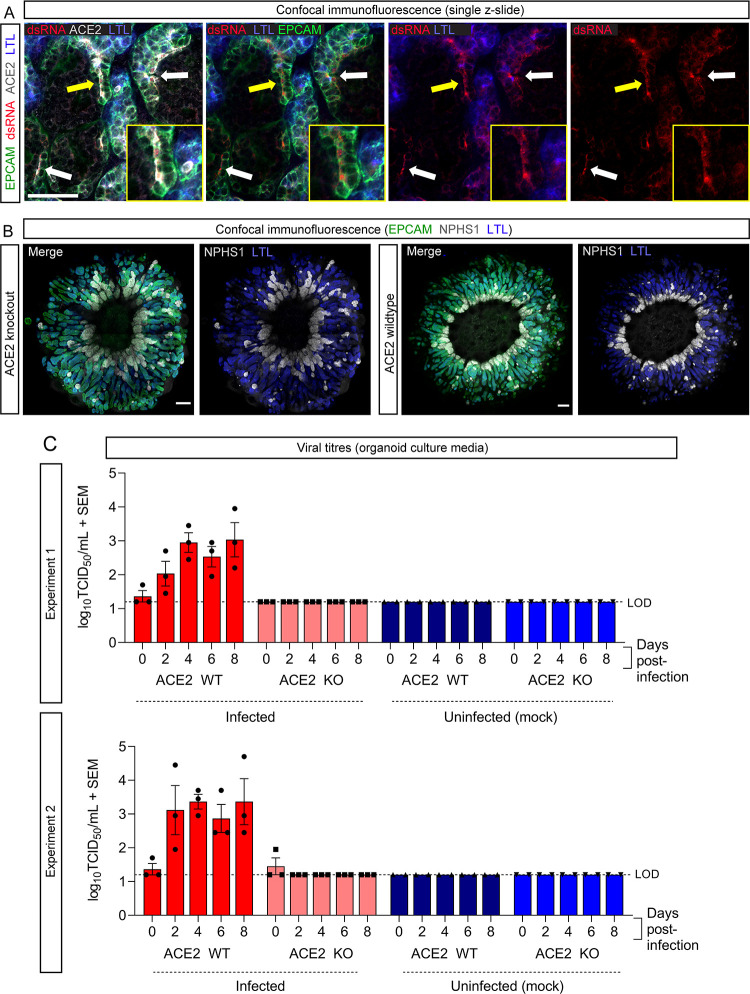
ACE2 is the sole receptor for SARS-CoV-2 in renal cells. (A) Confocal immunofluorescence of a representative PT-enhanced organoid 6 days post-infection demonstrating SARS-CoV-2 double-stranded RNA (dsRNA; red) localization within ACE2-positive (gray) proximal tubule cells. Organoid is co-stained for markers of proximal tubule brush-border membrane (LTL; blue) and nephron epithelium (EPCAM; green). Arrows indicate examples of dsRNA staining, with yellow arrow indicating the region shown at higher magnification in the inset images. Scale bar represents 50 µm. (B) Confocal immunofluorescence of PT-enhanced organoids (day 14 of organoid culture) generated from ACE2 knockout and wild-type iPSCs, depicting nephron epithelium (EPCAM; green), podocytes of the glomeruli (NPHS1; gray), and proximal tubules (LTL; blue). Scale bars represent 200 µm. Each confocal image depicts 3 × 3 stitched tiles, generated using the standard rectangular grid tile scan mode with automated stitching during image acquisition using ZEISS ZEN Black software (Zeiss Microscopy, Thornwood, NY) installed on a ZEISS LSM 780 confocal microscope (Carl Zeiss, Oberkochen, Germany). (C) Bar graphs from two independent experiments (top and bottom) depicting the viral titres (log10 TCID50/mL) of culture media sampled from ACE2 knockout (KO) and wild-type (WT) PT-enhanced organoids, both infected with VIC01 SARS-CoV-2 (dark and light red bars) or remaining uninfected (controls; light and dark blue bars). Error bars represent SEM from three biological replicates per timepoint. LOD, lower limit of detection.

