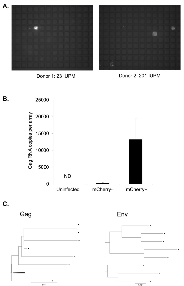
Fig 8.
Detection of HIV-infected cells from untreated PWH. (A) MOA cells were plated in CellRaft arrays at 4 cells per raft and then overlaid with PBMCs from untreated PWH at a density of 50 cells per raft. After 5 days of co-culture, the arrays were scanned, and mCherry-positive rafts were detected. (B) Sixty rafts were extracted from the arrays including mCherry-positive and mCherry-negative rafts, and RNA was isolated from each raft. The individual rafts were then quantified for HIV Gag RNA by qPCR. (C) Sequencing of partial Gag (HXB2 1195-1726) (left panel) and 3′ Half Genomes (HXB2 4923-9604) (right panel) RNA from virus from extracted mCherry rafts. Each sequence is represented by a circle. The reference bar indicates a phylogenetic distance of 0.001% nucleotide difference.

