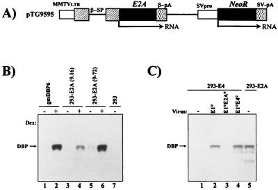
FIG. 1.
Structure of the E2A expression plasmid and steady-state levels of DBP protein in stable cell lines and after viral infection. (A) Schematic representation of the DBP expression plasmid pTG9595. Ad5 E2A sequences (nt 22440 to 24334) were inserted into an MMTV promoter-driven expression cassette (22) containing the rabbit β-globin splicing (β-SP) and polyadenylation (β-pA) signals. Expression of the neomycin resistance gene (NeoR) is regulated by the Simian virus 40 early promoter (SVpro) and late polyadenylation signal (SV-pA). LTR, long terminal repeat. (B) Western blot analysis of DBP protein in stable E1/E2A complementation cell lines. 293-E2A clones (9-16 and 9-72), established with pTG9595, and the control E2A complementation cell line gmDBP-6 (9) were compared for DBP expression in the presence (+) or absence (−) of dexamethasone (Dex). Total protein was extracted at 24 h postinduction, polypeptides (10 μg of protein) were separated on a 12% polyacrylamide–SDS gel, and the DBP protein was detected with the B6α72K anti-DBP monoclonal antibody (41) combined with enhanced chemiluminescence. (C) Comparison of DBP expression in 293-E2A cells (clone 9-72) and 293-E4 cells (clone 5-19; see Fig. 3) infected at an MOI of 6 IU/cell with AdE1°, AdE1°E2A°, and AdE1°E4° vectors. Total protein was extracted at 16 h p.i. (lanes 1 to 4) and 24 h after induction with dexamethasone (lane 5). DBP analysis was as described above.

