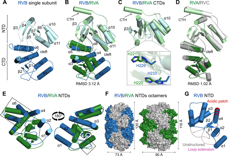
Fig 4.
Comparison of individual subunits of NSP2 from RVA, RVB, and RVC species of RVs. (A) RVB NSP2 subunit (present study). (B) Superposition of RVA NSP2 (PDB ID: 2R7C, green) on RVB NSP2 (present study, blue). (C) Superposition of CTDs of RVB and RVA NSP2s. A close-up view of catalytic histidine residues is shown in the inset. (D) Superposition of RVC NSP2 (PDB ID: 2GU0, gray) and RVA NSP2 (PDB ID: 2R7C, green). (E) Superposition of NTDs of RVB and RVA NSP2s (similar regions are indicated by rectangles). (F) View along the 2-fold symmetry axis of the NSP2 octamer with the NTD in blue (RVB) and green (RVA). (G) N-terminal domain of RVB NSP2 with unique features of the RVB clade marked as the following: unstructured region of RVB NSP2 in gray, conserved loop extension in pink, and acidic patch present in the RVB clade in red. In all panels, particular domains are indicated by a darker shade (NTDs) and lighter shade (CTDs) of each color. The C-terminal helix (CTH) is indicated by arrows.

