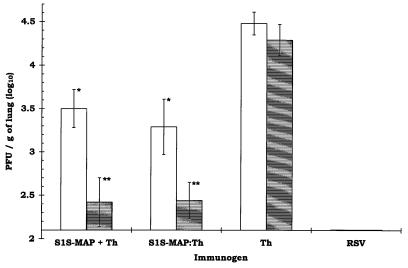
FIG. 1.
Reduction of RSV infection in the lungs of mice following immunization with S1S-MAP. The animals were challenged i.n. with RSV (106 PFU/50 μl) 4 to 5 weeks after the last boost. The lungs were removed 4 days later (at the peak of the viral load in the lungs) for quantification of viral titers. The results are expressed as log10 RSV PFU/g of lung tissue ± 1 standard deviation. The lowest level of virus detectable in this assay was 2.10 log10 PFU/g of lung tissue (12 PFU/animal). The animals in the RSV group (the positive control for protection) were infected with RSV (106 PFU/50 μl) 9 days prior to rechallenge with RSV. The open bars represent BALB/c mice (5 animals/group) immunized i.p. and boosted 3 weeks later with S1S-MAP plus Th, S1S-MAP–Th, or Th (5 nmol/animal). Anti-RSV IgG log10 titers for each group prior to challenge were as follows: S1S-MAP plus Th, 3.29 ± 0.13; S1S-MAP–Th, 3.01 ± 0.11; Th, <1.90. The shaded bars represent BALB/c mice (4 animals/group) immunized following the schedule described above but which received a second boost 6 weeks later. Anti-RSV IgG log10 titers for each group prior to challenge were as follows: S1S-MAP plus Th, 3.61 ± 0.20; S1S-MAP–Th, 3.38 ± 0.15; Th, <1.90. A significant reduction in RSV titers was observed in the lungs of all animals immunized with S1S-MAP plus Th and S1S-MAP–Th in comparison to that in the Th control group (∗, P < 0.01; ∗∗, P < 0.001 [Student t test]). Over five experiments, no significant differences were observed in RSV titers recovered from the lungs of Th-immunized and mock-immunized animals (P > 0.2 by one-way analysis of variance).

