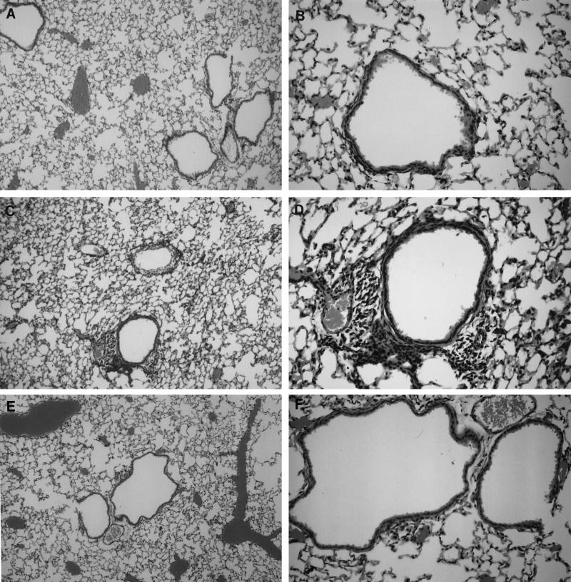
FIG. 2.
Sections of lung tissue stained with hemotoxylin and eosin showing reduced cellular infiltration in mice immunized with S1S-MAP plus Th constructs (A and B) compared to that in control mice immunized with Th alone (C and D) following challenge with RSV. As controls, sections from RSV-immunized and challenged mice showing no infiltration are also shown (E and F). The histological analyses were performed 7 days after challenge with RSV (106 PFU/50 μl). Magnification, ×100 (A, C, and E) and ×250 (B, D, and F).

