Fig. 2.
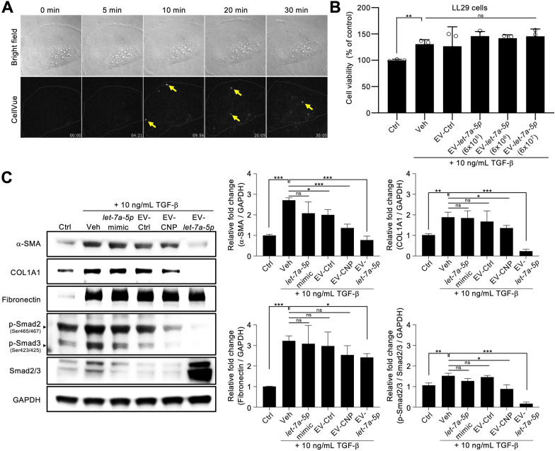
EV-let-7a-5p suppresses TGF-β-induced fibrotic phenotype in vitro. A Fluorescent images of stained cells were taken by Total Internal Reflection Fluorescence microscopy (TIRFM, Nikon) and quantitated for co-localization by Imaris software (BITPLANE, Oxford Instruments, Zurich, Switzerland). EVs were labeled with CellVue Claret and incubated with BEAS-2B cells at 37 °C for 30 min. B Cell viability was assessed using the SRB assay after 48 h of the indicated treatment. The cell density is 6000 cells per well treated with three concentrations of 6 × 10⁵, 6 × 10⁶, and 6 × 10⁷ EVs. C The fibrotic phenotype of LL29 cells was induced by TGF-β treatment. It demonstrated advanced fibrosis-related markers, characterized by increased Smad2/3 (phospho-Smad2/3) activation and elevated α-SMA, COL1A1, and fibronectin expression. Lipid nanoparticles, loaded with 10 pg of synthetic let-7a-5p and packaged using lipofectamine 3000, serve as a EV-let-7a-5p mimic (let-7a-5p mimic). This fibrotic response was effectively mitigated by EV-let-7a-5p (10,000 EVs per cell). Data were presented as mean ± standard deviation. The images are representative of n = 3 biologically independent experiments. For C, the data were analyzed by two-tailed unpaired Student’s t-test. (*p < 0.05, **p < 0.01, ***p < 0.001, ns = non-significant)

