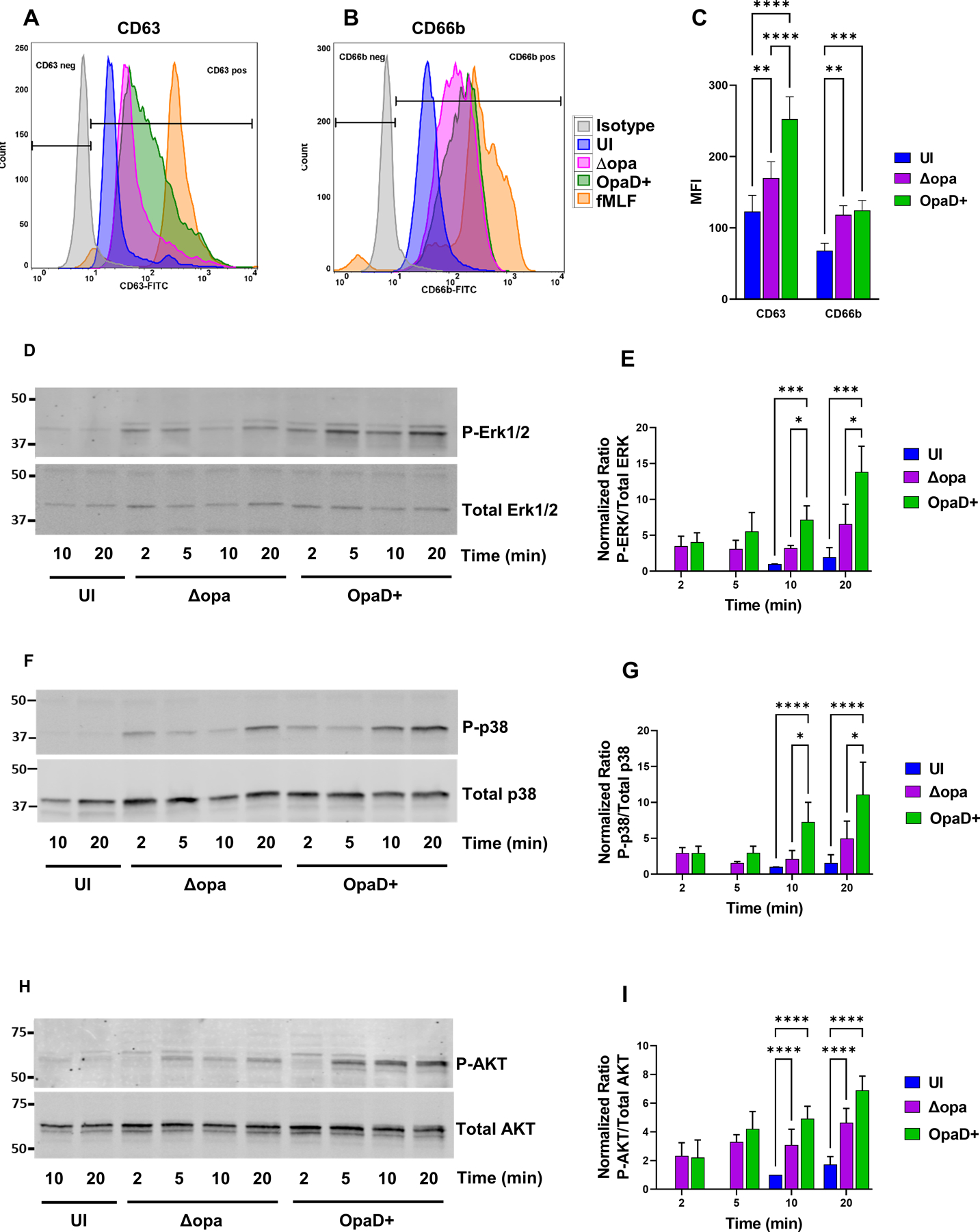
Figure 6. Limited activation of neutrophils following exposure to Opa-negative N. gonorrhoeae.

(A-C) Adherent, interleukin 8-treated primary human neutrophils were exposed to Δopa or OpaD+ Ngo for 1 hr, or left uninfected (UI). Surface presentation of CD63 (primary granules) (A) and CD66b (secondary granules) (B) was measured by flow cytometry. Neutrophils treated with cytochalasin B + fMLF served as a positive control for degranulation (fMLF). The geometric mean fluorescence intensity (MFI) ± SEM was calculated from n = 6 biological replicates (C), with statistical significance determined by two-way ANOVA with Sidak’s multiple comparisons test. (D-I) Adherent, IL-8 treated neutrophils were incubated with Δopa or OpaD+ Ngo or left uninfected for the indicated times (UI). Whole cell lysates were immunoblotted for phosphorylated and total p38, ERK1/2, and AKT. D, F, and H show a representative blot from one of three biological replicates. E, G, and I report the ratio of phosphorylated to total protein by quantitative immunoblot for the three replicates, relative to the uninfected condition at 10 min (whose ratio set to 1). Statistical significance was determined by two-way ANOVA followed by Tukey’s multiple comparisons test for 3 (G) or 4 (E, I) biological replicates. * P ≤ 0.05, ** P ≤ 0.01, *** P ≤ 0.001, **** P ≤ 0.0001.
